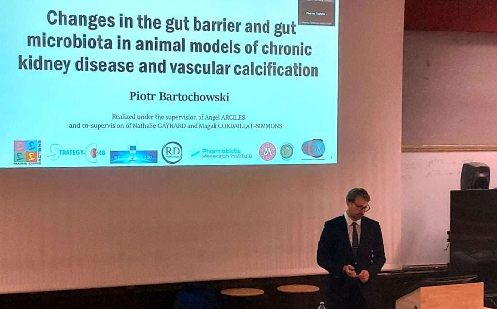
Piotr Bartochowski's thesis defense
Presented by Piotr Bartochowski
December 14, 2023
Edited by Àngel ARGILÉS,
And the co-supervision of Nathalie GAYRARD and Magali CORDAILLAT-SIMMON
Changes in the intestinal barrier and gut microbiota in animal models of chronic kidney disease and vascular calcification
Summary:
Chronic kidney disease (CKD) is characterized by a persistent reduction in renal function, accompanied by dysfunction of other organs. At the intestinal level, changes in the barrier and microbiota have been reported, which may contribute to disease progression through chronic inflammation of intestinal origin and the production of uremic toxins. In this context, the aim of this thesis is to identify changes in the intestinal barrier and microbiota during CKD.
In a literature review, the suitability of animal models of CKD for the study of the gut-kidney axis was investigated. Rodent models of CKD allow us to study changes in the gut and in the metabolism of the gut microbiota, but changes in the composition of the microbiota are not reproduced. The main factors affecting gut microbiota are the method of CKD induction, the animal species and housing conditions. To improve the transposition of results to humans, it is preferable to use omnivorous species whose CKD has been surgically induced and which are housed separately under controlled conditions.
The second study focused on characterizing the intestinal barrier of the ileum and colon in a rat model of CKD with or without vascular calcification (VC), using histological and molecular methods. VC is a complication of CKD characterized by the accumulation of phosphate and calcium crystals in arterial smooth muscle cells, inducing a loss of elasticity and contractility, which promotes cardiovascular disease. The main change observed in animals with CKD is a reduction in mucus production, which is greater in the presence of CVs. The presence of NLRP6, an important player in intestinal immunity, is reduced in the ileum of CKD animals with or without CV. NLRP6 gene expression is positively correlated with several gut genes related to tight junctions, immune response and the antioxidant system, and negatively correlated with renal fibrosis and plasma indoxyl sulfate levels. The concentration of this uremic toxin increases with CKD and is positively associated with CV and negatively with mucus production.
The third study aimed to identify changes in gut microbiota composition during CKD. These changes are assessed using metabarcoding, one of the next-generation sequencing methods. In renal failure rats, the composition of the gut microbiota does not appear to differ from the control group (overall composition, α-diversity and β-diversity). The few significant differences concern amplicon sequence variants with small contributions to the microbiota (including 13 unique to the CKD microbiota, 6 increased and 5 reduced compared with the control group). Significant differences in β-diversity were observed between the two points at baseline (before surgery) and at sacrifice. These results differ from the literature and are at least partly linked to methodological problems identified a posteriori.
In conclusion, animal models of CKD are well suited to the study of disturbances of the gut-kidney axis. For the first time, changes in the intestinal barrier in an animal model of CKD with CV have been reported. Reduced NLRP6 expression in the ileum suggests an important role in maintaining intestinal barrier homeostasis. A dysbiosis of the gut microbiota specific to CKD was not observed, but the study needs to be repeated with the inclusion of additional controls.
